
Focused Ion Beam Microscopy (FIB-SEM)
Focused Ion Beam Microscopy combines the methods of examining a sample using a focused ion beam (FIB) and an scanning electron microscope (SEM), allowing a sample to be examined and processed simultaneously.
Table of Contents
- How does focused ion beam microscopy work?
- Focused ion beam microscopy at Quality Analysis
- Preparation and 3D Analysis with FIB-SEM
- Examination methods in Focused Ion Beam Microscopy
- Advantages of Focused Ion Beam Microscopy
- Frequently asked questions about FIB-SEM
- Typical applications of FIB-SEM
- Summary: Focused Ion Beam Microscopy

Contact us –
expert support is just a conversation away.
Using state-of-the-art analytical methods, we assist you with quality issues, damage claims, and development challenges – providing precise, well-founded, and solution-oriented support.

How does Focused Ion Beam Microscopy work?
In FIB microscopy, an ion beam is focused using electromagnetic lenses and directed onto the sample. For analysis, the beam is guided across the surface, thereby scanning it. Either the emitted secondary electrons can be detected, or the intensity of the beam penetrating the sample. If, on the other hand, the FIB is to be used to ablate material, this is done in the same way, but with a significantly higher voltage (up to 50 kV).
Examination in a scanning electron microscope (SEM) works on the same principle as the FIB, but uses an electron beam generated by a field-emission cathode. In this method as well, secondary electrons can be detected for imaging. However, the contrast of the backscattered electrons is also frequently used, as it allows conclusions to be drawn about the different materials in a sample.
A FIB-SEM microscope combines both types of analysis, allowing the user to choose between them. It is also possible to use the ion beam to process the material and the electron beam to capture the image.
Focused Ion Beam Microscopy
at Quality Analysis
- High-resolution analysis of coatings, layer systems, microstructures and solder joints down to the nanometer scale
- Targeted preparation of cross-sections and material regions using focused ion beam (FIB) and femtosecond laser
- Investigation and visualization of defects, inclusions, impurities, and deposits at grain boundaries
- Analysis of intermetallic phases and other microstructures
- Determination of elemental composition using EDX analysis
- Layer thickness measurement and high-resolution evaluation of thin coating systems
- Rapid material ablation using a femtosecond laser to expose deeper regions
- 3D analysis through serial material removal, reconstruction, and defect detection
Preparation and 3D Analysis Using FIB-SEM
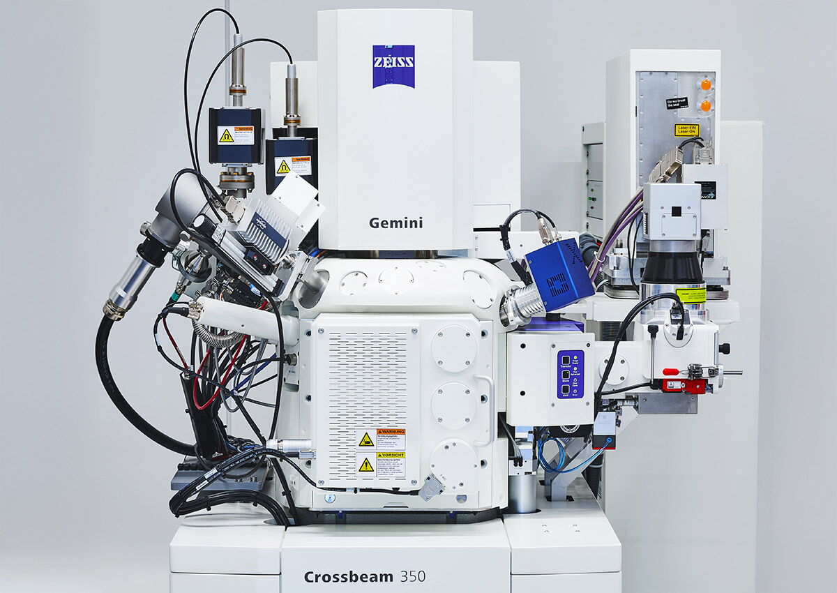
With the high-resolution ZEISS Crossbeam 350, we combine scanning electron microscopy, focused ion beam (FIB), and femtosecond laser technology. The FIB enables the precise preparation of minute material areas and cross-sections. With the femtosecond laser, larger volumes of material can be rapidly ablated to expose deeper regions, which can then be examined at high resolution.
Through serial material ablation and repeated imaging, three-dimensional volume structures can also be reconstructed. This enables not only high-resolution 2D cross-sections but also 3D analyses in the micro- and nanometer range—with minimal thermal and mechanical effects on the sample.
Examination methods in Focused Ion Beam Microscopy
A FIB-SEM microscope combines a focused ion beam system (FIB system) with a scanning electron microscope (SEM). Although imaging with both systems—including the focused ion beam system—is theoretically possible, in practice the ion beam is primarily used for sample preparation. The primary analytical instrument in focused ion beam microscopy is the electron microscope. Depending on the arrangement of the two systems, these are referred to as dual-beam or cross-beam microscopes.
Advantages of Focused Ion Beam Microscopy
Scanning electron microscopy is capable of producing high-resolution images of even the smallest samples; it is even possible to create three-dimensional images. However, to prevent the emitted electrons from interacting with atoms in the surrounding air, the examination must take place in a high vacuum. As a result, it is normally not possible to access the sample during the examination. This becomes a particular problem when a part of the sample is found to be of special interest only after examination in the SEM, or when precise material removal is required that can be performed reliably only under microscopic visual control.
The fine ion beam system solves this problem by allowing the processing of the sample using a second beam (with ions instead of electrons) during the examination. Since an FIB system operates on the same principle as a scanning electron microscope, it is relatively easy to combine the two systems. If the ion beam is to be used for processing rather than observation, the applied voltage must be increased. Another advantage is targeted material preparation with minimal thermal and mechanical effects. This allows even sensitive layer systems and the smallest material areas to be precisely exposed without unnecessarily affecting the structures under investigation.
Frequently asked questions about FIB-SEM
In scanning electron microscopy, the sample is imaged at high resolution using an electron beam. In FIB-SEM, a focused ion beam is also used to selectively ablate material. This makes it possible, for example, to create precise cross-sections and examine structures beneath the surface.
FIB-SEM is suitable for a wide range of materials and components, such as metals, ceramics, polymers, semiconductors, coatings, and electronic components. Depending on the research question, surfaces, coating systems, microstructures, interfaces, or localized defects can be examined. The ZEISS Crossbeam 350 is also designed for challenging or non-conductive samples.
Yes. When combined with EDX analysis, the elemental composition of selected areas can be determined. This allows for a more precise characterization of, for example, inclusions, impurities, deposits, or individual phases.
Typical Applications of FIB-SEM
Electronics and Microelectronics
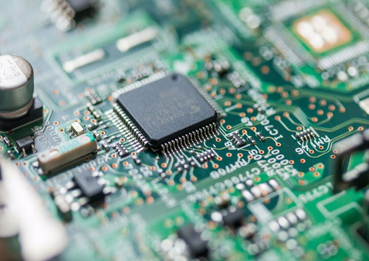
FIB-SEM can be used to examine printed circuit boards, solder joints, and electronic assemblies down to the nanometer scale. Targeted cross-sections reveal, for example, cracks, delaminations, and defects in conductive traces, metallization, or layer systems. In addition, semiconductor structures and material interfaces can be analyzed, layer thicknesses can be precisely determined, and microscopic defects can be identified.
Batteries and Fuel Cells
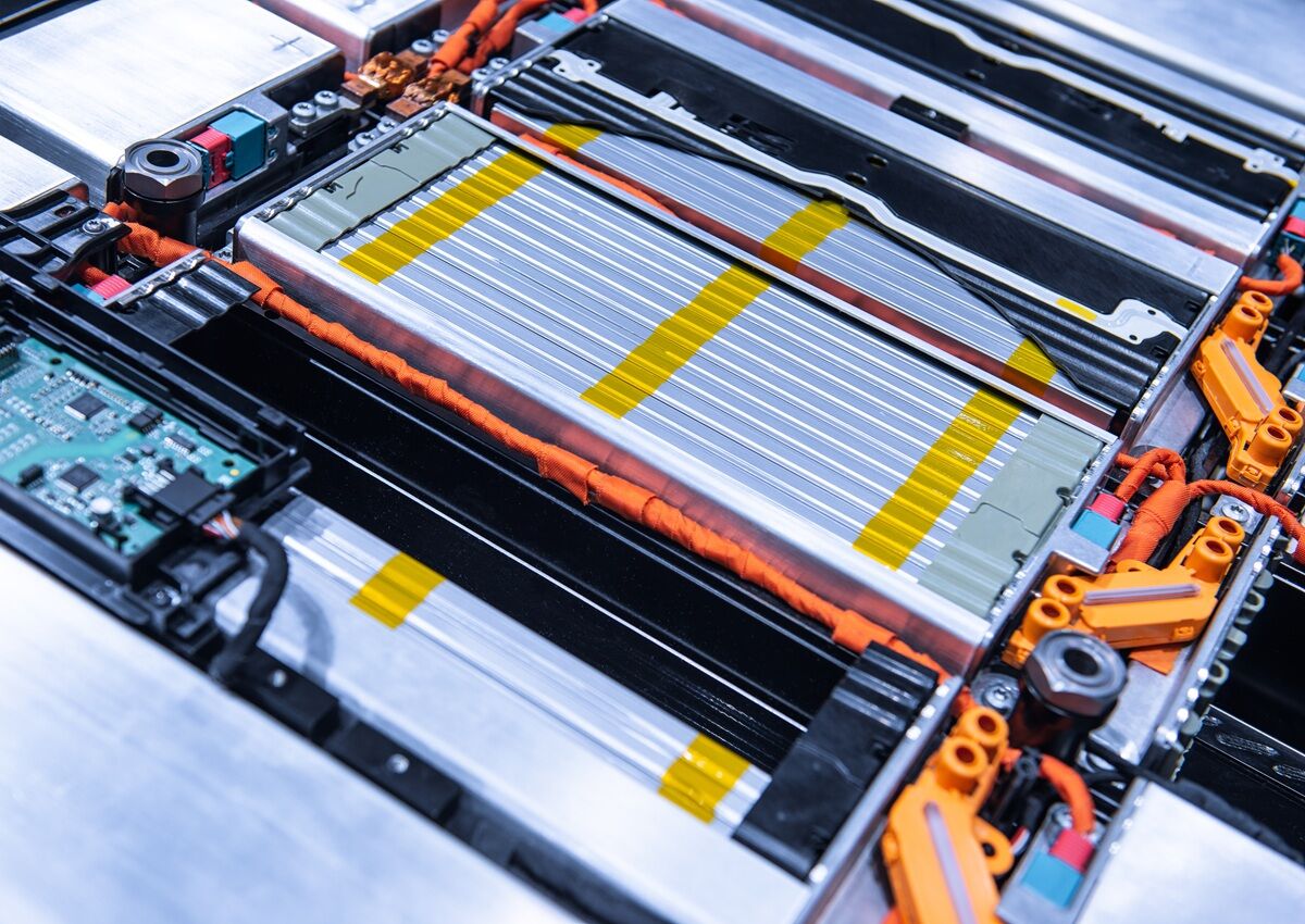
For batteries and fuel cells, FIB-SEM enables high-resolution analysis of electrodes, coatings, membranes, layer structures, and interfaces. Precise cross-sections aid in the evaluation of layer thicknesses and reveal local defects. In battery cells, for example, cracks, particle fractures, deposits, dendrites, and other signs of degradation can be examined in detail.
Coatings & Coating Systems
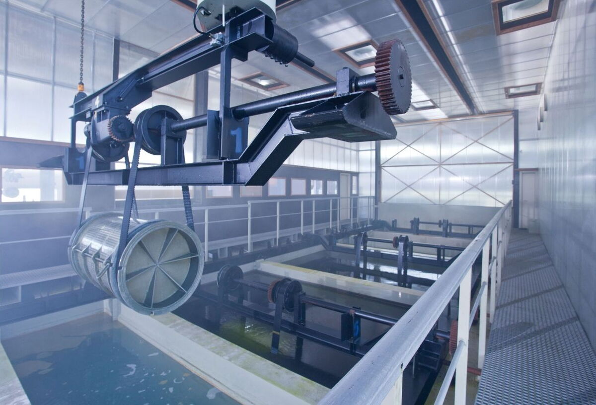
FIB-SEM enables targeted in-depth analysis of protective, functional, and multilayer systems. Targeted cross-sections provide information about layer structure, layer thickness, interfaces, and the bond to the substrate. At the same time, defects such as cracks, delaminations, pores, or inclusions can be detected, and individual material areas can be analyzed in detail.
Materials & Damage Analysis
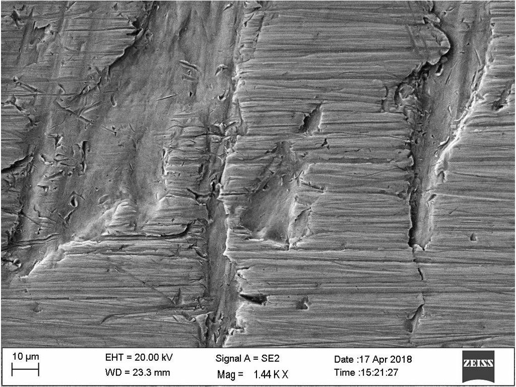
FIB-SEM allows specific local areas of material and microstructure to be exposed and examined down to the nanometer scale. This enables the analysis of, among other things, cracks, inclusions, impurities, pores, deposits at grain boundaries, and intermetallic phases. Precise cross-section preparation is particularly useful for examining microstructures and locally confined defects that are difficult to access using conventional methods.
Summary: Focused Ion Beam Microscopy
Focused Ion Beam Microscopy, or FIB-SEM for short, combines high-resolution scanning electron microscopy with a focused ion beam for targeted material preparation. This allows for the exposure of minute material areas, the creation of precise cross-sections, and the examination of structures down to the nanometer scale. Serial sectioning also enables three-dimensional analyses of microstructures and defects.